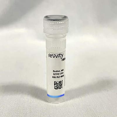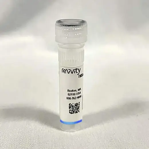
IVISense Bombesin Receptor 680 Fluorescent Probe (BombesinRSense)

IVISense Bombesin Receptor 680 Fluorescent Probe (BombesinRSense)


IVISense™ Bombesin Receptor 680 (BombesinRSense™ 680) is a targeted NIR fluorescent probe for in vivo imaging applications that targets bombesin receptors expressed in many types of cancers.
For research use only. Not for use in diagnostic procedures.
| Feature | Specification |
|---|---|
| Wave Length | 680 nm |
IVISense™ Bombesin Receptor 680 (BombesinRSense™ 680) is a targeted NIR fluorescent probe for in vivo imaging applications that targets bombesin receptors expressed in many types of cancers.
For research use only. Not for use in diagnostic procedures.


IVISense Bombesin Receptor 680 Fluorescent Probe (BombesinRSense)


IVISense Bombesin Receptor 680 Fluorescent Probe (BombesinRSense)


Product information
Overview
IVISense Bombesin Receptor 680 fluorescent probe is specific for bombesin receptors (BBRs) expressed in many types of cancer. Bombesin-like peptides and their G-protein coupled receptors have been shown to play a role in cancer and are overexpressed in a variety of tumors, including prostate, breast, lung, CNS, gastric, colon, and renal cell carcinomas.
IVISense Bombesin Receptor 680 imaging agent comprises a 7-amino acid bombesin peptide analog, an NIR fluorophore (ex/em 665/691 nm) and a pharmacokinetic modifier to improve its plasma availability (plasma t1/2 = 30-45 min). This agent can be used to target and quantify upregulation of bombesin receptors (BBR) in vivo associated with tumor proliferation.
Specifications
| Brand |
IVISense
|
|---|---|
| Fluorescent Agent Type |
Targeted
|
| Imaging Modality |
Fluorescence
|
| Shipping Conditions |
Shipped Ambient
|
| Therapeutic Area |
Oncology/Cancer
|
| Unit Size |
1 Vial (10 doses)
|
| Wave Length |
680 nm
|
Resources
Are you looking for resources, click on the resource type to explore further.
Fluorescence molecular imaging is the visualization of cellular and biological function in vivo to gain deeper insights into...
Researchers trust our in vivo imaging solutions to give them reliable, calibrated data that reveals pathway characterization and...
The goal of in vivo fluorescence molecular imaging is to enable non-invasive visualization and quantification of cellular and...
Epifluorescence (2D) imaging of superficially implanted mouse tumor xenograft models offers a fast and simple method for assessing...
In vivo fluorescence imaging displays a very broad utility and has become a well-established modality for functional imaging in...


How can we help you?
We are here to answer your questions.


