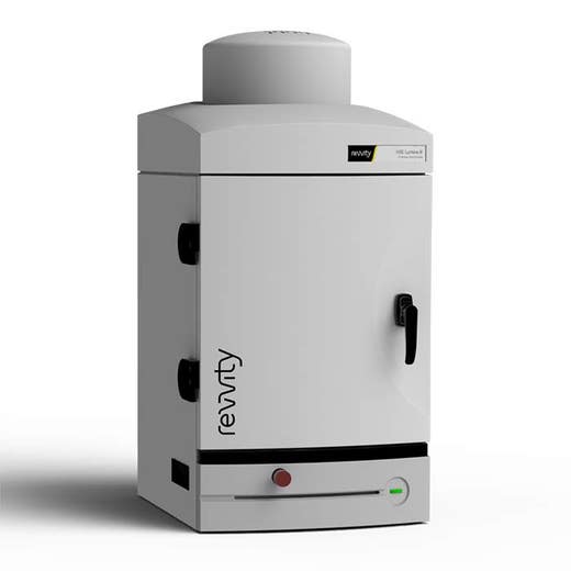
IVIS Lumina III In Vivo Imaging System


IVIS Lumina III In Vivo Imaging System


IVIS Lumina Series III benchtop 2D optical imaging system
IVIS Lumina III In Vivo Imaging System


The IVIS® Lumina Series III brings together years of leading optical imaging technologies into one easy to use and exquisitely sensitive bench-top system.
Product Variant
For research use only. Not for use in diagnostic procedures.
Product information
Overview
The IVIS Lumina III is capable of imaging both fluorescent and bioluminescent reporters. The system is equipped with up to 26 filter sets that can be used to image reporters that emit from green to near-infrared. Superior spectral unmixing can be achieved by Lumina III’s optional high resolution short cut off filters.
Features and Benefits
- Market trusted technology offering the fullest suite of leading imaging technologies, reagents and support
- Exquisite sensitivity in bioluminescence
- Full fluorescence tunability through the NIR spectrum
- Compute Pure Spectrum spectral umixing for ultimate fluorescence sensitivity
- Expandable system tailored to your workflow
- Complimentary Living Image™ software licenses are provided with the IVIS systems and upon request.
Specifications
| Dimensions | 48.0 cm (W) x 104.0 cm (H) |
|---|
| Brand |
IVIS
|
|---|---|
| Imaging Modality |
2D Bioluminescence
2D Fluorescence
|
| Unit Size |
1 Unit
|
References
- Lim et al (2021). Bioorthogonally surface‐edited extracellular vesicles based on metabolic glycoengineering for CD44‐mediated targeting of inflammatory diseases. J Extracellular Vesicles. https://doi.org/10.1002/jev2.12077
- Lee et al (2020). NIR dye-loaded mesoporous silica nanoparticles for a multifunctional theranostic platform: Visualization of tumor and ischemic lesions, and performance of photothermal therapy. J Indus. Eng. Chem. 88:99-105. https://doi.org/10.1016/j.jiec.2020.03.027
- Shim et al (2019). Carrier-free nanoparticles of cathepsin B-cleavable peptide-conjugated doxorubicin prodrug for cancer targeting therapy. Jrnl. Controlled Release. 294: 378-389. https://doi.org/10.1016/j.jconrel.2018.11.032
- Lee et al (2019). Crushed Gold Shell Nanoparticles Labeled with Radioactive Iodine as a Theranostic Nanoplatform for Macrophage-Mediated Photothermal Therapy. Nano-Micro Letters. 11(36). https://doi.org/10.1007/s40820-019-0266-0
- Zhang et al (2019). In vivo irreversible albumin-binding near-infrared dye conjugate as a naked-eye and fluorescence dual-mode imaging agent for lymph node tumor metastasis diagnosis. Biomaterials. 217:119279. https://doi.org/10.1016/j.biomaterials.2019.119279
- Singh et al (2019). A Novel Orally Active Inverse Agonist of Estrogen-related Receptor Gamma (ERRγ), DN200434, A Booster of NIS in Anaplastic Thyroid Cancer. Clin Cancer Res. https://doi.org/10.1158/1078-0432.CCR-18-3007
- Sonoda et al (2018). A Blood-Brain-Barrier-Penetrating Anti-human Transferrin Receptor Antibody Fusion Protein for Neuronopathic Mucopolysaccharidosis II. Mol Ther. 26(2): 1366-1374. https://doi.org/10.1016/j.ymthe.2018.02.032
- He et al (2018). Repurposing disulfiram for cancer therapy via targeted nanotechnology through enhanced tumor mass penetration and disassembly. Acta Biomater. 28: 113-124. https://doi.org/10.1016/j.actbio.2017.12.023
- Jung et al (2017). Hydrophobically modified polysaccharide-based on polysialic acid nanoparticles as carriers for anticancer drugs. Int. J Pharmaceutics. 520(1-2): 111-118. https://doi.org/10.1016/j.ijpharm.2017.01.055
- Kafa et al (2016). Translocation of LRP1 targeted carbon nanotubes of different diameters across the blood–brain barrier in vitro and in vivo. Jrnl. Controlled Release. 225: 217-229. https://doi.org/10.1016/j.jconrel.2016.01.031
Resources
Are you looking for resources, click on the resource type to explore further.
Technical Note
Living Image software to draw measurement ROIs in 2D and 3D optical images taken on IVIS systems
Technical Note
Subtracting background ROI from the measurement ROIs when performing fluorescence quantitative analysis


How can we help you?
We are here to answer your questions.


