
IVIS Lumina S5 Imaging System


IVIS Lumina S5 Imaging System


IVIS Lumina S5 Imaging System


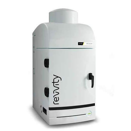
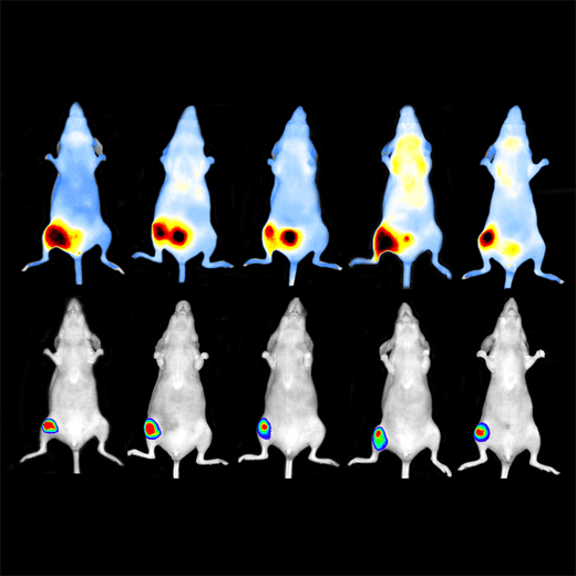
The IVIS® Lumina™ S5 high-throughput 2D optical imaging system combines high-sensitivity bioluminescence and fluorescence in a benchtop format. With an expanded 5 mouse field of view for 2D optical imaging plus our unique line of accessories to accelerate setup and labeling, it has never been easier or faster to get robust data- and answers- on anatomical and molecular aspects of disease.
For research use only. Not for use in diagnostic procedures.
Product information
Overview
The IVIS Lumina S5 in vivo imaging system has all the capabilities of the current IVIS Lumina platform with improved throughput and accessories to streamline imaging workflow, data acquisition and analysis, ideal for accelerating your research.
High-throughput High-Sensitivity Optical Imaging
The IVIS Lumina S5 integrates a 1 inch CCD camera into our benchtop instrument providing a high throughput 20 x 20 cm Field of View (FOV) sufficient for imaging 5 animals at a time for bioluminescence and fluorescence in vivo imaging.
As with other IVIS Lumina in vivo optical imaging systems, the S5 is equipped with 26 filters tunable to image fluorescent sources that emit from green to near-infrared. Novel illumination technology effectively increases fluorescent transmission through 900 nm. Moreover, the IVIS Lumina S5 incorporate Revvity's patented Compute Pure Spectrum (CPS) algorithm for spectral library generation software tools to ensure accurate autofluorescence removal, unmixing and fluorophore quantitation. Standard on all IVIS instruments, absolute calibration affords consistent and reproducible results independent of magnification, filter selection from one instrument to any another IVIS instrument within an organization or around the world.
IVIS Lumina S5 – A High Throughput Solution
Not only does the IVIS Lumina S5 offer higher throughput via the 1 inch CCD, but it is also compatible with a set of smart animal handling accessories (purchased separately) designed with throughput and safety in mind.
Smart loading trays will allow users to pose animals on the benchtop before placing the tray into the IVIS. Fiducials built into the tray will allow the software to automatically recognize and draw ROIs providing automated animal identification.
Animal trays are designed with ease of use and user safety in mind. No nose cones are required thus minimizing cleanup. All tray parts are autoclaveable for ease of sterilization and when used with the next generation anesthesia unit (RAS-4), strong vacuum capabilities minimize excess gas from escaping thus preventing exposure of users to anesthetic gas.
Finally, Living Image® software brings IVIS technology to life by facilitating an intuitive workflow for in vivo optical image acquisition, analysis and data organization. The software’s design creates an intuitive, seamless workflow for researchers of all skill levels. Living Image will support input of unique animal IDs when using chip technologies and readers from third party vendors thus streamlining labeling, setup and subsequent export of data for analysis.
Key Features:
- High throughput (5 mouse) optical imaging
- Supports mouse and rat imaging
- Compute Pure Spectrum (CPS) spectral unmixing
- Full fluorescence tunability through the NIR spectrum
- Unique accessories to speed acquisition and analysis
- Small footprint–sits on your benchtop
- Complimentary Living Image™ software licenses are provided with the IVIS systems and upon request.
Specifications
| Height |
106.68 cm
|
|---|---|
| Width |
48.26 cm
|
| Brand |
IVIS
|
|---|---|
| Imaging Modality |
2D Bioluminescence
2D Fluorescence
|
| Unit Size |
1 Unit
|
Video gallery


IVIS Lumina S5 Imaging System


IVIS Lumina S5 Imaging System


References
- Fichman et al (2021). Plasmodesmata-localized proteins and ROS orchestrate light-induced rapid systemic signaling in Arabidopsis. Science Signalling. 14(671). https://doi.org/10.1126/scisignal.abf0322
- Zhuang et al (2020). mRNA Vaccines Encoding the HA Protein of Influenza A H1N1 Virus Delivered by Cationic Lipid Nanoparticles Induce Protective Immune Responses in Mice. Vaccines (Basel). 8(1). https://www.ncbi.nlm.nih.gov/pmc/articles/PMC7157730/
- Fink et al (2020). Loss of Ing3 Expression Results in Growth Retardation and Embryonic Death. Cancers. 12(1). https://www.mdpi.com/2072-6694/12/1/80#
- Shong et al (2020). Serotonin Regulates De Novo Lipogenesis in Adipose Tissues through Serotonin Receptor 2A. Endocrinology & Metabolism. 35(2): 470-479. https://doi.org/10.3803/EnM.2020.35.2.470
- Fink et al (2019). Capacity of the medullary cavity of tibia and femur for intra-bone marrow transplantation in mice. PLOS ONE. https://doi.org/10.1371/journal.pone.0224576
- Zhao et al (2018). Hyaluronic Acid Layer-By-Layer (LbL) Nanoparticles for Synergistic Chemo-Phototherapy. Pharmaceutical Res. 35(196). https://doi.org/10.1007/s11095-018-2480-8
Resources
Are you looking for resources, click on the resource type to explore further.
Optical imaging detects photons in the visible range of the electromagnetic spectrum. PET and SPECT imaging instruments are...
Aside from the traditional small-molecule chemotherapeutics or targeted therapy agents that have been widely used in the clinic...


How can we help you?
We are here to answer your questions.


