Cellometer Auto T4 Brightfield Cell Counter
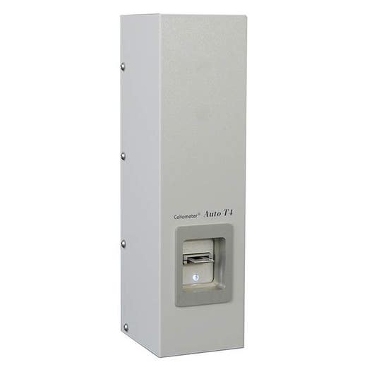

Cellometer Auto T4 Brightfield Cell Counter

Cellometer Auto T4 Brightfield Cell Counter


The Cellometer™ Auto T4 automated cell counter is designed for concentration and Trypan blue cell viability assessment of cell lines, even clumpy cells, with optional GMP/GLP software.
For research use only. Not for use in diagnostic procedures.
Product information
Overview
The Cellometer Auto T4 utilizes brightfield imaging and pattern-recognition software to quickly and accurately identify and count individual cells. Cell count, concentration, diameter, and % viability are automatically calculated and reported.
The Cellometer Auto T4 cell counter offers:
- On screen view of individual cell level information
- Ability to capture multiple fields of view for accurate data
- Built-in cell type library
- Count difficult cells (clumpy, irregular-shaped)
- Options for IQ/OQ Validation and GMP/GLP software
Additional product information
One-step concentration & viability
The Cellometer Auto T4 simultaneously calculates cell concentration and % viability for cultured cells stained with trypan blue.
Trypan blue viability is a dye exclusion method that utilizes membrane integrity to identify dead cells. The dye is unable to penetrate healthy cells, so they remain unstained. Dead cells have a compromised cell membrane that is permeable to the trypan blue dye. Dead cells are stained blue and display as dark cells in the Cellometer software with brightfield imaging.

Within 10 seconds, the Cellometer Auto T4 instrument reports:
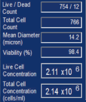
- Live, dead, and total cell count
- Live and total cell concentration
- Mean Cell Diameter
- % Viability
Counted brightfield image

Live cells = green circle
Dead (stained) cells = red circle
Multiple cell images for data verification
With the Cellometer Auto T4 Cell Counter, cell morphology can be immediately viewed on-screen.
Two images with four fields of view each are captured per cell count. This is equivalent to the area of four quadrants of a hemocytometer. Counted cells are indicated on-screen for further verification that cells in the sample are being imaged and analyzed properly.
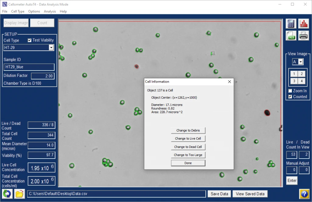
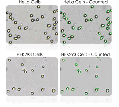
Users can confirm that:
- Cells are counted correctly, based on size and shape
- Debris is excluded
- Cells within clumps are being counted individually
- Cell images can be archived and exported for use in publications and presentations
- Saved images can be re-counted using default or user-optimized analysis settings
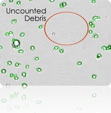
Exclusion of debris
The Cellometer distinguishes cells from cellular debris based on size, brightness, and morphology. This allows for increased accuracy with cell counts.
- Cell size parameters can be modified to optimize exclusion of debris from results and enhance the accuracy of counting for a wide range of cell sizes.
- The counted image can be viewed to verify exclusion of debris from results.
Cellometer precision
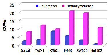
The Cellometer %CV (Coefficient of Variation), including sample preparation, was well under 5% for most cell lines tested. The CVs for manual counts ranged from 7% to 20%. Automated cell counters remove inter-operator variability and subjectivity from cell counts to generate more precise data.
Improve data accuracy & consistency
- Eliminate wash steps
- Reduce judgment errors
- Reduce recording & calculation errors
- Reduce counting time... Run more experiments
| Cellometer Auto T4 | Hemacytometer | ||||
|---|---|---|---|---|---|
| Concentration | n | CV | Concentration | n | CV |
| 1.17 x 10⁶ | 4 | 2.27% | 1.11 x 10⁶ | 3 | 8.04% |
| 1.36 x 10⁶ | 4 | 3.17% | 1.41 x 10⁶ | 3 | 11.89% |
| 6.36 x 10⁵ | 4 | 8.29% | 5.58 x 10⁵ | 3 | 9.66% |
| 1.32 x 10⁶ | 4 | 5.57% | 1.16 x 10⁶ | 3 | 20.65% |
| 3.81 x 10⁵ | 4 | 2.79% | 3.70 x 10⁵ | 3 | 19.49% |
| 9.18 x 10⁵ | 4 | 0.39% | 7.68 x 10⁵ | 3 | 10.60% |
Cellometer Auto T4 brightfield cell counter specifications
| Includes |
|
|---|---|
| Available accessories |
|
| Imaging performance |
|
| Instrument specifications |
|
| PC / Laptop requirements: (If purchasing Cellometer without PC laptop) |
|
Specifications
| Brand |
Cellometer
|
|---|---|
| Unit Size |
1 Each
|
Resources
Are you looking for resources, click on the resource type to explore further.


How can we help you?
We are here to answer your questions.


